The future in tinnitus treatment
Home

Call us on +61 8 8333 1010


Ten Important Management Strategies
________________________________
Apply these simple techniques to manage your tinnitus and reduce your level of tinnitus perception.
Call us on +61 8 8333 1010


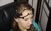
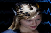
Several central nervous system phenomena have been proposed to explain the emergence of tinnitus. In individuals with tinnitus, changes in the electroencephalograph (EEG) are pronounced in recordings from temporal regions which strongly correlate with the level of tinnitus related perception and distress.
Research also suggests that in tinnitus the midbrain auditory nuclei receive substantial sensory innervation from higher centres and the subjective experience of sensory phenomena is strongly modulated by attentional affects which can cause changes in auditory cortex activity levels.
Modulation of these parameters can be achieved through the specialised application of either one or a combination of tinnitus counselling, neurofeedback and/or hypnosis.
Home

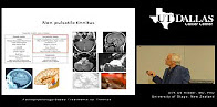
Many different functional and structural imaging techniques have been used to identify structures in the central nervous system which are believed to play an important role in the pathophysiology of many forms of tinnitus.
The neuroimaging methods functional magnetic resonance (fMRI) and positron emission
tomography (PET) enable to measure regional changes of cerebral blood flow, which
in turn is an indirect measurement of neuronal activity. Electro-
EEG and MEG have revealed consistent results across many studies in the sense that in tinnitus the normal activity pattern in the auditory cortex is changed. In the auditory cortex of tinnitus patients alpha activity is reduced, whereas delta and gamma activity is increased. Successful treatment reverses these abnormalities, indicating that they represent the neuronal correlate of tinnitus loudness.
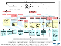
TRI Protocols -
This flowchart is an interactive PDF-
You find detailed information by clicking the boxes of the main flowchart slide. Links to explanatory slides are always marked by an arrow. By clicking on the "home" symbol in the bottom right corner you get easily back to the main slide.This TRI Flowchart for Patient Management will contribute to a better diagnosis and treatment of the many tinnitus patients worldwide,